Enhancing Surgical Preoperative Planning Through VR-Based 3D Organ Visualization
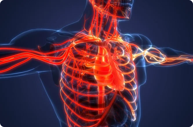
The assurance of successful surgery depends on surgeons being able to establish spatial connections between anatomical and pathological structures mentally. Additionally, preoperative planning of surgery procedures highly depends on computer assistance. Various planning software solutions were introduced to support surgeons in preoperative planning. Surgeons use these to mentally reconstruct the internal representation of a body part and build spatial relationships with the particular patient. This task can be challenging even for well-trained surgeons. A 3D organ visualization can provide substantial support for such actions. Several possibilities allow for obtaining a 3D model with detailed inner structures, vascular structures, and tumors from the tomographic data.

Examining a 3D model using a regular 2D screen loses the advantages of 3D display methods because they cannot convey depth cues such as binocular disparity and motion parallax.
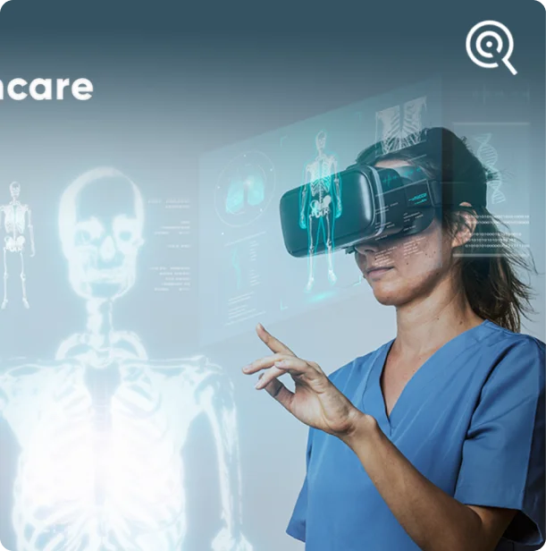
In contrast, a VR system mimics how we perceive the physical world. With head-mounted displays (HMD), the user gets the impression of seeing natural 3D objects. Therefore, VR has proved effective for numerous surgical simulations, including training fundamental surgical skills.
At Otto-von-Guericke University, they presented a multi-user conference room prototype for surgery planning inside VR, where users can benefit from interaction with 3D organ models and 2D gray-value images. This system also enables the discussion of surgical problems over distance. They chose liver surgery planning for evaluation purposes, but this prototype is also functional for planning other surgical procedures. In an initial investigation, surgeons found this tool valuable for preoperative planning procedures. You can have access to this study through link bellow.

©2022 Implident. All Rights Reserved.
